


 e140 860

lined by an endocervical mucinous-type epithelium,
surrounded by normal or mildly hyperplastic smooth
muscle. The lumen contains mucus with neutrophils.
Cytological atypia and gland crowding are absent
[9]
.
The differential diagnosis includes urachal remnants,
cystitis glandularis (intestinal type), primary adenocarcino-
ma, and metastatic adenocarcinoma
[4,5]
. The urachal
remnants are usually found in the anterior or apical aspect
of the bladder dome, and not in the posterior aspect as for
endocervicosis. They form a tortuous line representing a
single canal lined by mucinous epithelium surrounded by
fibrous tissue, which in turn is associated with layers of
smooth muscle cells. The epithelial lining does not contain
ciliated cells, endometrioid glands, or endometrial-type
stroma. In the intestinal type of cystitis glandularis, the
glands are typically located in the subepithelial connective
tissue and do not involve the detrusor. In addition, the glands
can be associated with a typical component of cystitis
glandularis and lack endometrioid glands or endometrioid/
elastotic-type stroma, features indicative of mu¨ llerian origin.
Primary adenocarcinoma of the bladder, originating from
the surface epithelium, may show a deceptively benign
microscopic appearance. However, the glands are present
throughout the bladder wall, with prevalent involvement of
the mucosa, and are often associated with a urothelial cell
component. The cells show at least focally greater atypia,
and the glands have angulated ends and a desmoplastic
stromal response. Some endocervical adenocarcinomas may
have secondary involvement of the urinary bladder wall and
show a deceptively benign appearance such as in adenoma
malignum. In such cases the patient should have other
clinical symptoms suggestive of the cervical origin of the
tumor. A primary adenocarcinoma can arise on a background
of endocervicosis. In such cases there is a transition from
benign glands to dysplastic to franklymalignant glands
[4,5]
.
4.
Endosalpingiosis and mu¨ llerianosis
Endosalpingiosis is a benign mu¨ llerian lesion with glands
lined by ciliated cells similar to the tubal epithelium
[4,5,10]
. Occasionally, endosalpingiosis is seen in a pure
form. However, more often it is associated with an
endocervical and endometrioid-like mu¨ llerian glandular
epithelium
( Fig. 2 ). This mixture of benign epithelial
[(Fig._1)TD$FIG]
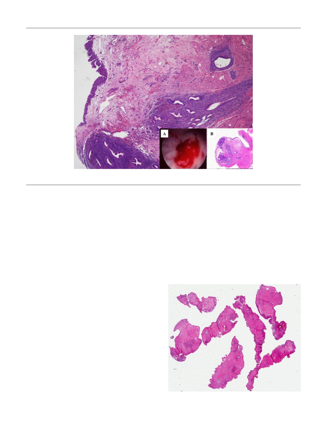
Fig. 1 – Endometriosis involving the bladder mucosa. Inserts: (A) cystoscopic appearance and (B) low-power histology.
[(Fig._2)TD$FIG]
Fig. 2 – Low-power histology of Mu¨llerianosis. A high-resolution version
of this slide for use with the Virtual Microscope is available as eSlide
VM04124.
E U R O P E A N U R O L O G Y 7 2 ( 2 0 1 7 ) e 1 3 9 – e 1 4 1
e140